



2The orofacial myofunctional functions and structures6
3Orthodontic clinical situations related to orofacial dysfunctions17
Introduction. Contemporary orthodontics integrates orofacial functions that actively participate in craniofacial growth, sleep quality, attention and behavior, and therapeutic stability. Beyond dental alignment, the clinical challenge is twofold: early detection of dysfunctions (nasal vs oral breathing, tongue posture/swallowing, mastication, speech) and their correction through a hierarchical and reproducible sequence.
Materials and methods. Narrative review based on a selection of clinical studies, systematic reviews, expert consensus, and international clinical recommendations, focusing on the prevalence, pathophysiology, and management of orofacial myofunctional dysfunctions in orthodontics.
Results. Seven orofacial functions and structures are detailed: lingual frenulum (≈8% prevalence of ankyloglossia, with a doubled overall risk of malocclusion), breathing (robust associations between mouth breathing, maxillary transverse deficiency, hyperdivergent Class II, and anterior open bite), parafunctions (digit sucking correlated with open bite and posterior crossbite), cranio-cervical posture (bidirectional relationships between cervical posture and sagittal skeletal class), mastication (habitual unilateral chewing associated with 73% condylar asymmetries vs 21% in controls), tongue posture/swallowing (dysfunctional swallowing perpetuating malocclusions), and speech (strong correlations between articulation disorders and open bite/Class II–III). A controlled study shows that combining orthodontics with orofacial myofunctional therapy significantly reduces relapse of anterior open bite (63% without relapse vs 0% with orthodontics alone; mean relapse 0.48 mm vs 3.38 mm; p < 0.0001). This article presents the SPF pyramid (Structure–Posture–Function), a reappraised functional framework that integrates all elements of myofunctional therapy within orthodontic care and hierarchizes management into three steps: (i) removal of obstacles; (ii) correction of the functional environment; (iii) active myofunctional therapy.
Discussion. The gold standard pathway relies on referral to specialized myofunctional therapists. Passive myofunctional appliances can complement but not replace active myofunctional therapy based on neuroplasticity principles (Kleim & Jones 10 principles) and biofeedback. Digital tools offer opportunities to improve access to care and documentation.
Conclusion. Systematic integration of functional screening and orofacial myofunctional therapy into the orthodontic pathway, according to a hierarchical sequence (breathing, frenulum, parafunctions, posture, swallowing, mastication, speech), improves therapeutic stability and justifies structured interdisciplinary coordination.
Keywords: Orthodontics / Myofunctional Therapy / Mouth Breathing / Malocclusion / Tongue Position / Ankyloglossia / Nasal Breathing / Craniofacial Growth / Functional Orthodontics / Orofacial Dysfunctions
1Introduction
Contemporary orthodontics is part of a medical approach in which orofacial myofunctional processes actively contribute to craniofacial growth, to the proper course of orthodontic treatment, and to the stability of its results. Beyond dental alignment, the clinical challenge is twofold: to screen for dysfunctions early and to correct them within a hierarchical, reproducible sequence (16, 20, 63). The high prevalence of orofacial myofunctional disorders in the primary and mixed dentitions — with, for example, dysfunctional swallowing in 62–63.5% of children and the presence of at least one orofacial myofunctional disorder in nearly 9 out of 10 (88.8% in primary dentition and 89.8% in early mixed dentition) — justifies integrating functional management alongside orthodontic treatment (83, 32). In the pediatric orthodontic population, approximately 10.8% of children present a positive risk of obstructive sleep-disordered breathing, nearly double that of the general pediatric population (1). Historically, the link between orofacial functions, growth, and orthodontic stability has long been established, with Rogers describing as early as 1939 the principles of a myofunctional therapy grounded in orofacial muscular balance (77).
Objective — To offer a critical narrative review of recent data and to present an operational clinical framework, the SPF pyramid (Structure–Posture–Function), for structuring the management of orofacial myofunctional dysfunctions in orthodontics according to a hierarchical sequence (screening, removal of obstacles, active myofunctional therapy), and to strengthen its therapeutic stability as well as interdisciplinary coordination.
1.1 Professional consensus and international recommendations
The assessment of orofacial functions is progressively becoming a recognized component of the diagnosis and management of orthodontic treatment. Most international clinical recommendations and learned societies now recognize that a comprehensive orthodontic examination must include a functional assessment (breathing, swallowing, lingual frenulum and tongue posture, speech, mastication, parafunctions) — and that the clinician must be able to demonstrate that it was performed (16, 20, 63, 94). These same recommendations advocate the early correction of identified disorders. Specifically regarding obstructive sleep-disordered breathing, the White Paper of the American Association of Orthodontists confers on the orthodontist a screening role — history-taking, clinical examination, and a validated questionnaire — before referral to the sleep physician (8), a position reaffirmed in its update (67). Far from being an isolated requirement specific to one country, this orientation is the subject of an international convergence that positions functional screening integrated into the orthodontic work-up as a recognized clinical standard, directly linked to the quality and stability of treatments. It justifies documentation of screening, measurements, and referrals, as well as a multidisciplinary and protocol-based organization of care.
1.2 Guiding principles (neurofunctional and biomechanical)
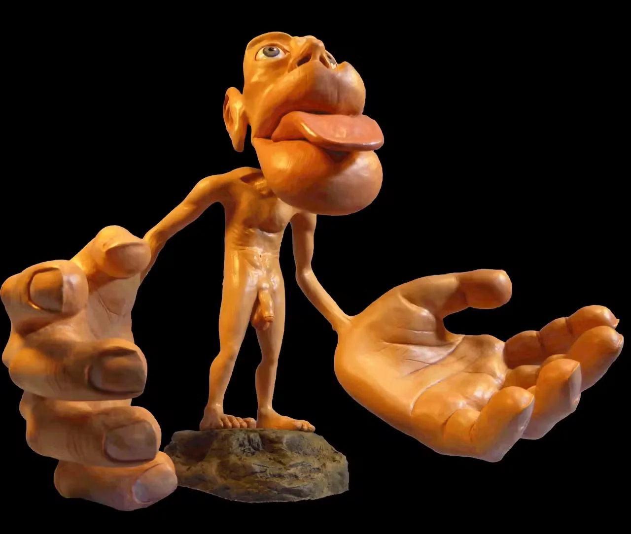
The over-representation of the tongue, lips, and perioral region in Penfield’s sensorimotor homunculus underscores the proprioceptive density and motor learning capacity of the orofacial sphere (69). The functional matrix theory (Moss) posits that craniofacial morphogenesis is largely determined by the activity of functions and adjacent soft tissues; in practice, a tongue against the palate stimulates transverse maxillary growth and the development of the nasal cavity, whereas persistent mouth breathing lowers the tongue, reduces palatal stimulation, and sustains the dentofacial deformity (65). Wolff’s law reminds us that bone, a living anisotropic material, adapts to mechanical stresses: the absence of physiological pressures (e.g., a low tongue posture that prevents the repeated micropressures of the tongue against the palate) favors narrow, high-vaulted palatal architectures (90). These principles provide the biological and neurofunctional foundation of orofacial myofunctional therapy.
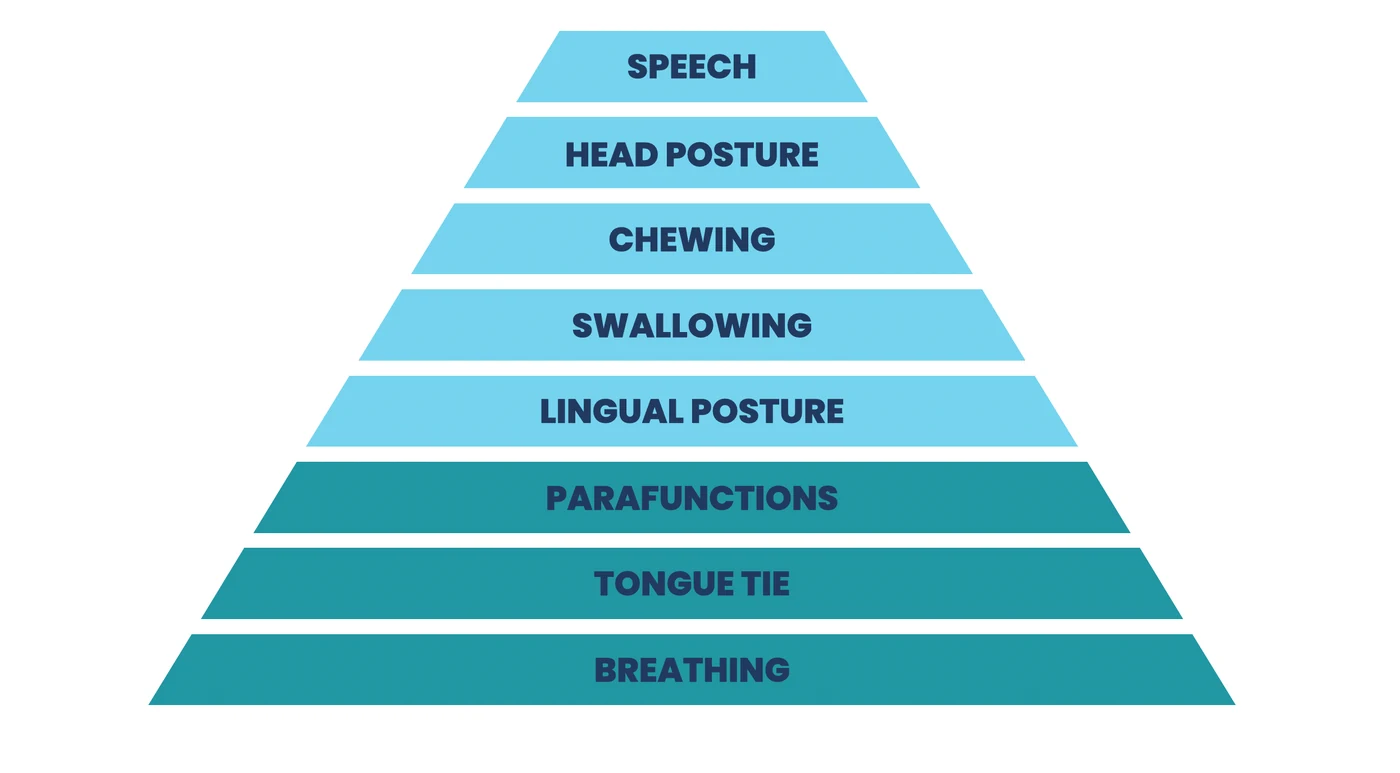
1.3 Functional pyramid (hierarchical logic of management)
In this article, we propose a reappraised functional pyramid — the SPF pyramid (Structure–Posture–Function) — which formalizes an operative hierarchy integrating, beyond the orofacial functions, the structural and postural determinants relevant to myofunctional therapy applied to orthodontics.
- S — Structures: restrictive lingual frenulum; nasopharyngeal obstacles affecting patency.
- P — Postures: resting tongue posture and cervico-cephalic posture.
- F — Functions:
- Nasal breathing: restore patency and the automatization of closed-mouth breathing.
- Parafunctions: eliminate interferences.
- Resting tongue posture & swallowing: retrain palatal contact and swallowing patterns without parasitic contractions.
- Mastication: aim for chewing on alternating sides (alternating unilateral mastication).
- Global posture: integrate the cervico-cephalic postural determinants.
- Speech: retrain articulatory patterns.
This organization translates into an operative three-stage timeline, whose stages may intertwine while respecting the guiding order:
- Removal of obstacles: restore nasal breathing, treat a restrictive lingual frenulum when present, and wean off parafunctions.
- Correction of the functional environment: within the framework of orthodontic treatment.
- Active myofunctional therapy: train and automatize the orofacial myofunctional functions within the new environment.
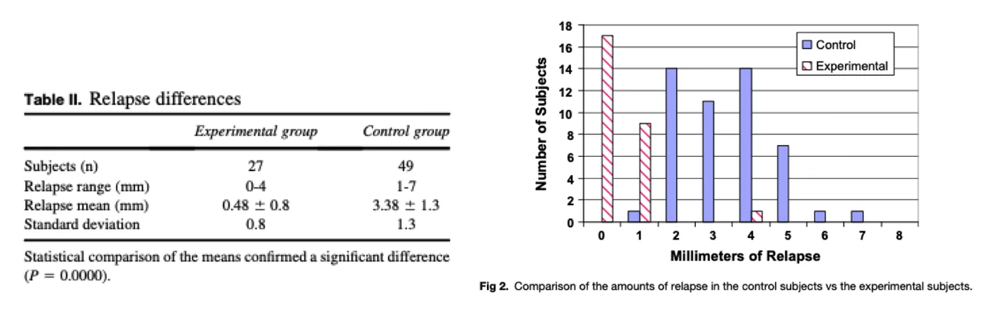
The clinical relevance of integrating orofacial myofunctional therapy into orthodontic treatments is illustrated by the comparative study of Smithpeter & Covell: in 76 patients with anterior open bite, combining orthodontics + orofacial myofunctional therapy (an individualized program of up to 49 exercises, ≈ 14 sessions) significantly reduced relapse compared with orthodontics alone; 63% of subjects in the myofunctional group showed no relapse versus 0% in the control group, and the magnitude of relapses was markedly lower (0.48 ± 0.8 mm vs 3.38 ± 1.3 mm; p < 0.0001) (81). This clinical evidence, combined with the high prevalence of dysfunctions in the general pediatric population (83), reinforces the need to integrate orofacial myofunctional therapy into orthodontic treatments (Figure 3).
This issue is not limited to children. In adults, orofacial myofunctional dysfunctions remain factors of delay and complications in the course of orthodontic treatments, and constitute a recognized factor of relapse after the end of treatment, the persistence of a neuromuscular imbalance opposing the stability of the result obtained (58, 41, 80).
2The orofacial myofunctional functions and structures
2.1 Lingual frenulum
Ankyloglossia is a variation in the embryological development of the frenulum that limits the physiological mobility of the tongue. A meta-analysis of 24,536 infants (< 1 year) reports a prevalence of approximately 8%, with a male predominance close to 2:1 (p < 0.01) (38). Beyond the sole difficulty of suck-swallow in the infant, several studies associate ankyloglossia with dento-skeletal disorders: a multicenter, matched, blinded study conducted in 100 children shows that a short frenulum doubles the overall risk of malocclusion, with a strengthened link for Class III (p = 0.029) (9). In a multicenter cohort of 700 children, frenula classified as Kotlow III–V are correlated with anterior open bite, a narrow maxilla, and lower incisor diastemas (p < 0.05), whereas very short frenula are less represented in Class I–II (86). Finally, regarding stability, adding orofacial myofunctional therapy to orthodontics significantly improves the durability of the results (cf. Smithpeter & Covell, above) (81).
Recent studies confirm that lingual frenectomy, when combined with orofacial myofunctional therapy, significantly improves tongue mobility, resting tongue posture, and the quality of orofacial functions, with a favorable impact on swallowing, speech, and orthodontic stability. Tecco et al. (2015) showed that combining surgery + myofunctional therapy leads to a faster normalization of swallowing patterns and better elevation compared with surgery alone (84). These results are corroborated by Zaghi et al. (2019, 2023), who report, in large cohorts, an improvement in nasal breathing, posture, and tongue function in > 80% of cases after frenuloplasty combined with myotherapy, with an excellent safety profile (92, 93). Baxter et al. (2020) emphasize that this integrated approach is part of a modern functional understanding of the restrictive frenulum, considered as an embryological remnant limiting the physiological mobility of the tongue (6). In an orthodontic context, Nammour et al. (2019) indicate that frenectomy, particularly laser-assisted, favors optimal tongue positioning, supports nasal breathing, and may contribute to transverse maxillary growth by removing anterior mechanical constraints (66). Lichnowska et al. (2021) finally show that articulatory recovery depends closely on post-surgical myofunctional therapy (53). On the biomechanical and postural level, Rocabado (1983) and Giannakopoulos et al. (2013) recall the relationships between the tongue, hyoid bone, and cervico-cephalic posture, suggesting that releasing the frenulum may contribute to better craniofacial musculoskeletal balance (76, 29). Overall, the literature supports that frenectomy is an anatomical lever allowing orofacial myofunctional therapy to fully express its efficacy when interdisciplinary coordination is respected (84, 6).
2.1.1 Definition and physiology
Ankyloglossia refers to a congenitally short, thick, or fibrous lingual frenulum that limits the elevation, protraction, and sometimes the lateral movement of the tongue (38). The International Affiliation of Tongue-Tie Professionals more recently agreed on the definition of the restrictive lingual frenulum, defining it as an “embryological remnant of tissue in the midline between the undersurface of the tongue and the floor of the mouth that restricts normal tongue movement” (6). This definition addresses ankyloglossia from a more functional standpoint because it highlights the aspect of limited mobility without the frenulum necessarily appearing abnormal visually.
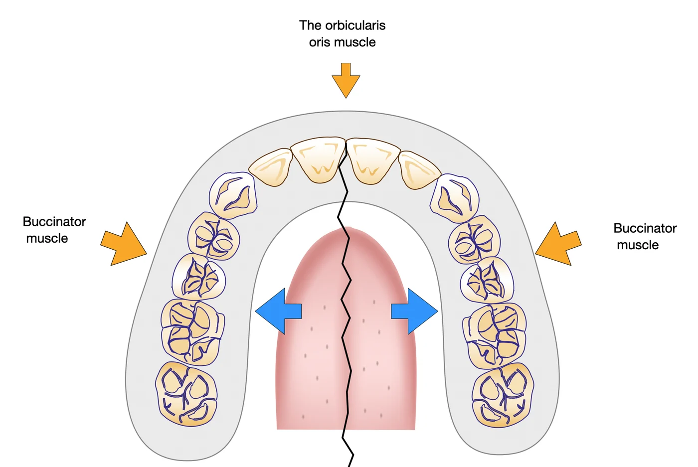
On the physiological level, the architecture of the arches results from a muscular balance. The position of the teeth stabilizes within a neutral zone — historically described as Château’s “dental corridor” or Danguy’s “zone 0” — where the centripetal forces (lateral buccinator muscles, orbicularis oris anteriorly) and the centrifugal forces exerted by the tongue at rest and during functions are balanced. A restrictive frenulum limits the palatal contact of the tongue, alters its high resting posture, and modifies this balance, predisposing to morphological consequences.
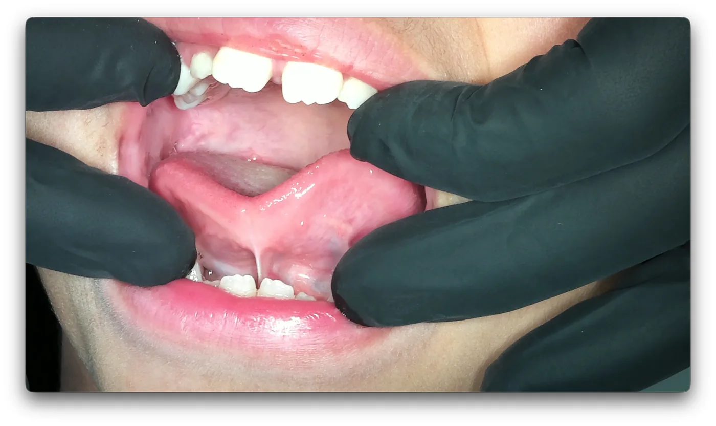
2.1.2 Pathophysiology and dysmorpho-functional cascade
The repercussions affect all 3 dimensions:
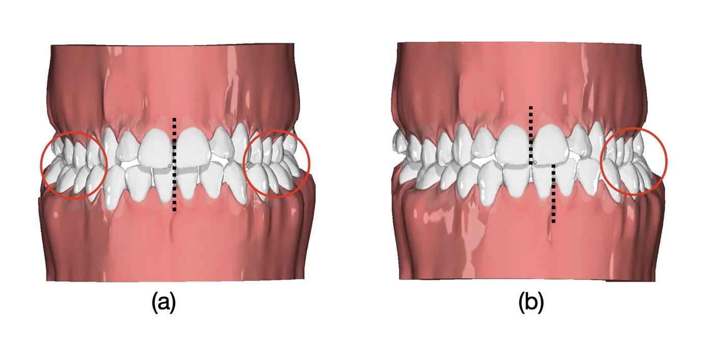
- Transverse: when tongue elevation is hindered, the tongue remains low and anterior. The decrease in palatal pressures allows the forces of the cheeks and lips to predominate: the neutral zone migrates inward, leading to a maxillary/alveolar constriction that clinically favors crossbites. In seeking maximum intercuspation, the mandible may deviate on closing (functional lateral deviation), with a shift of the incisor midlines; maintained during growth, this kinetic anomaly then evolves into a skeletal anomaly (laterognathia).
- Anteroposterior: a narrow upper arch behaves like a transverse lock. In certain patients, the mandible projects forward to interlock, producing an anterior crossbite (functional Class III) — consistent with the reported association between a short frenulum and a Class III phenotype (9). Conversely, the lock may mask a lateral crossbite, through a mandibular retroposition: this is referred to as relative maxillary alveolar constriction, which is revealed transversally when the patient is positioned in an Angle Class I relationship.
- Vertical: the tongue interposed during swallowing prevents incisor contact and sustains the anterior open bite, with labial version of the incisors from repeated pressures (86).
2.1.3 Screening and measurements
The examiner will assess 3 determinants during examination of the frenulum:
- texture;
- insertion;
- impairment of tongue functionality.
Screening combines clinical examination and simple functional tests: asking the patient to touch the retro-incisive papillae without mandibular compensation, at maximum mouth opening (Wilder and Gelesko test). A frenulum is considered restrictive when the amount of mouth opening with the apex of the tongue on the retro-incisive papillae is less than half of the patient’s maximum mouth opening. The Kotlow classification is based on measuring the distance between the tip of the tongue and the anterior attachment of the frenulum when the tongue is raised. It allows estimation of the free length of the tongue, considered as an indicator of the severity of a restrictive frenulum (48).
2.1.4 Management
The strategy is multidisciplinary and hierarchical. As a first-line approach, when mobility is partially preserved, stretching exercises are favored. When the frenulum is too restrictive, a frenectomy is indicated. Two points condition the functional prognosis:
- Preoperative: the patient must become familiar with the exercises before the intervention in order to master the technique according to the anatomical possibilities.
- Immediate postoperative: the early resumption of exercises (within the hours following the procedure) is essential to prevent fibrous healing/re-attachment, failing which the gain in mobility is lost and the discomfort may persist or worsen. This is then referred to as a scar band.
Management combines, depending on the case, the dental surgeon/ENT-maxillofacial surgeon, the therapist, and the orthodontist. Frenectomy alone is not sufficient: anchoring the benefits requires orofacial myofunctional therapy immediately after the procedure.
2.2 Breathing
2.2.1 Definition and physiology
Nasal breathing is the physiological ventilatory route in humans. It provides three inseparable functions — filtration, warming, and humidification — which condition the quality of tracheobronchial air and ventilatory expenditure. As air passes through the nasal cavities, it is trapped by the vibrissae and mucus and then swept along by the ciliated epithelium, eliminating the majority of particles larger than 5 µm. The inferior and middle turbinates act as a thermal and hygrometric exchanger: inspired air is brought to ~33–34 °C and its relative humidity approaches 80–98 %, parameters that are essential for efficient gas exchange. Compared with mouth breathing, nasal breathing achieves equivalent oxygen consumption for a minute ventilation reduced by 35 to 38%, typically accompanied by a lower respiratory rate and a slightly increased tidal volume, reflecting better ventilatory efficiency during exercise (18, 22). This superiority is due in particular to paranasal nitric oxide production, whose vasodilatory and anti-infectious effects optimize alveolar diffusion and pulmonary perfusion.
The “nasal cycle” — the alternation every two to six hours of one dominant nostril and then the other — results from neurovegetative tonic variations of the turbinates and must be known in order to correctly interpret patency tests. Finally, nasal breathing has a morphogenic dimension: the daily passage of 12,000 to 15,000 L of air exerts repeated stresses on the nasal walls and the floor of the nasal cavity, which is mechanically continuous with the palatal vault; within the logic of the functional matrix, this stimulation is thought to contribute to the transverse growth of the midfacial complex (65).
Several observational surveys link mouth breathing to malocclusions. In 1,616 preschool children, Paolantonio et al. demonstrated a significant association between mouth breathing, posterior crossbite, moderate overjet, and anterior open bite. In a cohort of 128 mouth-breathing subjects, Cozza et al. reported an increased prevalence of Class II, hyperdivergence, and anterior open bite. Souki et al., in 401 children assessed in a specialized ENT clinic, confirmed the high frequency of posterior crossbite (≈ 30 % in the mixed dentition and ≈ 48 % in the permanent dentition) among mouth breathers. Harvold’s experimental primate model — nasal obstruction with silicone plugs — reproduces an increase in lower facial height, a low tongue posture, and a downward and backward mandible, bringing the phenotype closer to clinical hyperdivergence. These observational and experimental studies converge on a robust association between chronic mouth breathing, transverse maxillary deficiency, a Class II pattern from mandibular rotation, and anterior open bite (68, 17, 82, 37, 54).
2.2.2 Pathophysiology and the dysmorpho-functional cascade
Prolonged mouth breathing develops on a dual background:
- an obstructive factor promotes mouth breathing;
- lip incompetence and/or postural habits perpetuate the open-mouth posture.
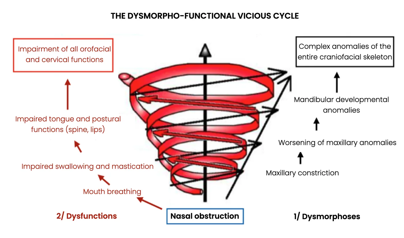
The tongue, forced to abandon its resting palatal posture to let air pass, ceases to stimulate the maxillary sutures laterally: the neutral zone is altered, resulting in a transverse deficiency. This transverse lock hampers mandibular advancement and promotes a rotation (hyperdivergent Class II) through the mandibular position in mouth breathing. In parallel, lingual interposition during functions maintains the anterior open bite. Together, this illustrates a bidirectional dysmorpho-functional cascade in which the skeletal and functional components are self-perpetuating (17, 37, 65, 68, 82).
2.2.3 Screening and diagnosis
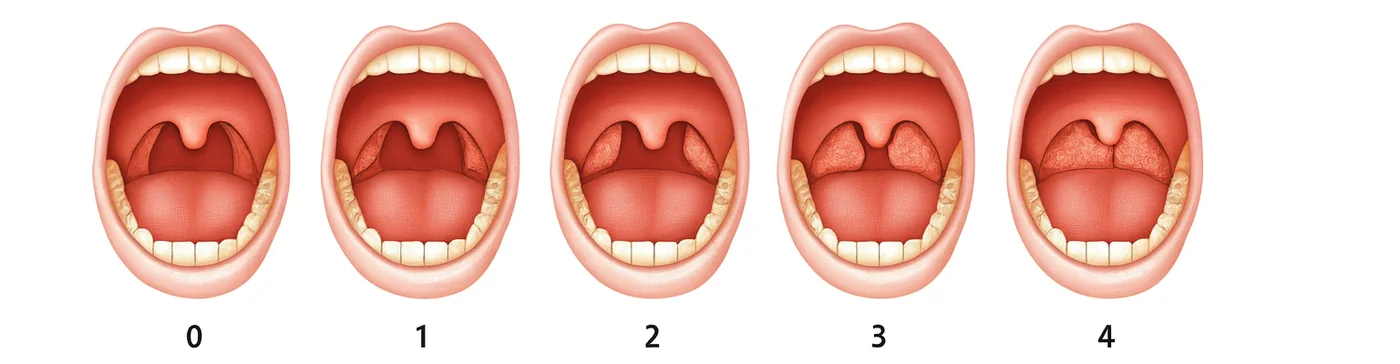
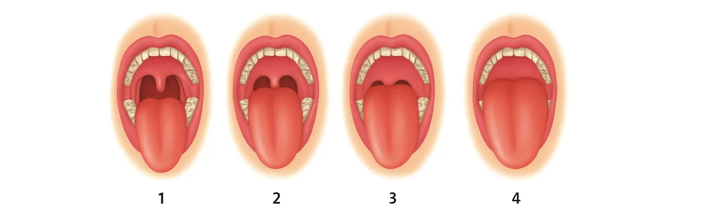
The Glatzel test (symmetrical condensation of mist on a cold metal mirror held beneath the nostrils) provides a simple assessment of patency, to be interpreted in light of the nasal cycle. The Gudin test reveals nasal valve incompetence that may or may not be related to an anatomical obstruction. The Rosenthal test assesses the patient’s ability to maintain strict nasal breathing over 15 cycles, thus reflecting their functional capacity to breathe through the nose. The extraoral examination looks for the stigmata of the adenoid facies (infraorbital dark circles, thin nostrils, a slightly open mouth, dry lips, a hyperdivergent typology, drooping palpebral fissures); the intraoral examination assesses tongue posture and lip competence. Oropharyngeal evaluation can be objectified using the Friedman and modified Mallampati scores (27, 57).
2.2.4 Orthodontic and craniofacial repercussions
In the transverse dimension, the most consistent consequence is maxillary constriction, often associated with posterior crossbites (17, 68, 82). The resulting mandibular functional shift illustrates the functional genesis of asymmetries liable to become skeletal. In the anteroposterior dimension, the transverse lock promotes Class II through mandibular rotation; in the vertical dimension, hyperdivergence and anterior open bite reflect tongue lowering and persistent open-mouth posture (17, 37, 82). Narrow upper arches increase the risk of eruption anomalies, in particular canine impaction.
2.2.5 Management: principles and protocols
Treatment is sequential and interdisciplinary. It begins with the removal of obstacles (ENT / allergist, by surgery or local treatment) and then with the normalization of volumes. Increasing the transverse maxillary dimension (surgical or orthopedic) increases the volume of the nasal cavities and reduces resistance to flow; Poiseuille’s law explains why small transverse gains can produce a major respiratory benefit (18, 22). Consolidation relies on orofacial myofunctional therapy.
Furthermore, if there is no anatomical obstacle, myofunctional therapy can begin in the case of nasal/labial/buccal immaturity. The latter will rely on proprioceptive and postural work, together with advice on nose-blowing and nasal hygiene.
2.2.6 Functional genioplasty
In some patients, passive lip closure remains impossible without contraction of the mentalis muscle despite the absence of an anatomical obstacle and despite myofunctional therapy. In these situations, functional genioplasty is a brief surgical procedure that modifies soft-tissue support, restores lip competence, and facilitates nasal breathing, indirectly improving swallowing, speech, and orthodontic stability, particularly when vertical excess of the lower third or a skeletal Class II persists. Indicated when resting mouth breathing and lip incompetence persist, it benefits from pubertal timing: performed around the pubertal growth spurt, prospective series report mandibular redirection (increased SNB, anterior rotation) compared with later interventions (25). In adolescents with obstructive breathing disorders, a return of nasal breathing, spontaneous lip closure, and improvement in nocturnal symptoms are observed after genioplasty (26). Other studies describe a gain in retrolingual space and better oropharyngeal patency (7), without a deleterious effect on growth and with more pronounced bone remodeling in younger patients (12). A systematic review reports an average increase in the posterior pharyngeal space of approximately 4 mm and, in several series, volumetric gains in the upper airway after genioplasty (30). The procedure is not a substitute: it creates a favorable environment that must be consolidated by orofacial myofunctional therapy.
2.2.7 Sleep apnea: reference points for the orthodontist
Obstructive sleep apnea-hypopnea syndrome (OSA) is defined by the repetition of episodes of partial or complete obstruction of the upper airway during sleep, inducing desaturations and micro-arousals. The apnea-hypopnea index (AHI) qualifies severity (adult: ≥5/h, ≥15/h, ≥30/h; child: ≥1/h, ≥5/h, ≥10/h). The disease is common and largely underdiagnosed (14, 44). It affects approximately 1 to 5 % of children in the general population (57), but nearly 10.8 % in the pediatric orthodontic population (1). This over-representation is most likely explained by the link between the deformities frequently seen in these patients (maxillary constriction, retromandibulism, hyperdivergence) and the obstructive risk (15). In children, it is accompanied by attention disorders, decreased academic performance, and, owing to the fragmentation of deep sleep, growth retardation (31). In adults, it increases cardiovascular mortality, daytime sleepiness, and the risk of accidents. The predominant collapse sites of the upper airway are the soft palate (~84 %) and the base of the tongue (~52 %) (51), hence the importance of myofunctional therapy aimed at toning these structures in the absence of a neuromuscular etiology (central apneas). The etiologies are divided among:
- anatomical factors (adenoids/tonsils, macroglossia, epiglottic collapse, septal deviation, retromandibulism, maxillary constriction, overweight);
- functional factors (hypotonia of the oropharyngeal muscles, nasal valve incompetence, mouth-breathing habits);
- positional factors (supine position);
- and other, rarer causes (acromegaly, craniofacial anomalies…).
Any clinical suspicion warrants referral and a sleep assessment. Indeed, assessment of the craniofacial phenotype alone is unreliable for identifying pediatric OSA, the effect of facial characteristics being modulated by age and body mass index (23); in children, screening therefore relies on a validated questionnaire such as the PSQ (14). In adults, dedicated tools such as the STOP-BANG and the Epworth scale are used (44).
Recent meta-analyses confirm the value of orofacial myofunctional therapy in the management of obstructive sleep apnea (OSA), in adults as well as in children. The work of Camacho et al. (2023), Xie et al. (2022), Hu et al. (2025), and Cammaroto et al. (2024) shows that myofunctional therapy, focused on strengthening the lingual, velar, and perioral muscles, reduces the apnea-hypopnea index (AHI) by an average of 30 to 50 %, improves minimum oxygen saturation, and decreases daytime sleepiness (10, 11, 39, 91, 34, 72). In adults, it optimizes the efficacy and adherence of conventional treatments (CPAP, mandibular advancement device). In children, it contributes to the restoration of nasal breathing and to the correction of the often-associated orofacial myofunctional dysfunctions. From this perspective, Cohen-Lévy et al. (2023) emphasize that myofunctional therapy constitutes an indispensable therapeutic adjunct in the multidisciplinary management of pediatric OSA, by reinforcing nasal breathing and the stability of orthodontic results. By acting on the tone and coordination of the oropharyngeal structures, myofunctional therapy appears to be an essential complementary etiological approach (15). As an illustration, a randomized controlled trial showed that daily didgeridoo training, toning the tongue and soft palate, reduced daytime sleepiness and the severity of OSA (74).
2.3 Parafunctions
Oral parafunctions comprise habits that serve no physiological purpose (nutrition, speech, swallowing) and that, when repeated or prolonged, alter the neuromuscular environment and the balance of forces applied to the arches. The most common are non-nutritive sucking (thumb, finger, pacifier, lip), object biting (nails, pens, “skin”) and, more broadly, certain masticatory parafunctions (bruxism). Their morpho-functional impact is explained by a disruption of the balance of the forces present within the neutral zone: interposition of the finger or pacifier prevents lingual elevation against the palate and reduces transverse stimulation (centrifugal forces), while sucking hyperactivates the buccinator, a constrictor muscle, and increases the centripetal forces on the neutral zone. Through pressure and the “uncapping” movement, the thumb induces labial version of the maxillary incisors and deepens the palate. It also promotes linguoversion of the lower incisors and hampers mandibular growth. Digital interposition maintains an anterior open bite and a low tongue posture, with associated dysfunctional swallowing and phonetic disorders.
Several clinical studies confirm the association between parafunctions and malocclusions. In 444 children in the mixed dentition, Warren et al. reported links between non-nutritive sucking and the occurrence of anterior open bite as well as posterior crossbites (89). In a population of 265 subjects, Cozza et al. showed that a sucking habit is associated with maxillary narrowness and an increased prevalence of posterior crossbite, particularly in hyperdivergent phenotypes (p < 0.01) (17). Finally, in a cohort of 1,099 children, Viggiano et al. demonstrated a significant association between parafunctions and posterior crossbite (p < 0.0001) (87).
2.3.1 Functional consequences and the dysmorpho-functional cascade
Thumb sucking establishes dysfunctional swallowing by interposition and a low tongue posture at rest, which maintains mouth breathing and disrupts chewing and speech. Over the long term, these disturbances fit into a dysmorpho-functional cascade: the habit (sucking) induces a dysfunction (low tongue, dysfunctional swallowing) that fuels maxillary developmental anomalies (transverse deficiency) and, by continuity, hinders mandibular growth, with a global repercussion on craniofacial balance.
2.3.2 Screening and principles of management
Screening relies on questioning the patient and the parents if the patient is a child, on the extraoral/intraoral examination, and on the assessment of functions.
Management is educational and behavioral: clear information for the child and the parents, empowerment and reinforcement, substitution strategies. Simple aids may be used (bitter varnish, anti-thumb gloves) when adherence is present.
Coercive devices (anti-thumb grids, glued spurs) are not recommended: they address neither the behavioral intention nor the motivation and can be circumvented, at the cost of adverse effects (unwanted movements on the anchor teeth). Psychological support is indicated if the habit persists into adolescence/adulthood, or coexists with significant anxiety.
2.4 Posture
2.4.1 Definition and pathophysiological framework
Posture results from a multimodal integration (visual, vestibular, somatosensory) within a craniocervicomandibular continuum. The afferents and efferents of the cervicofacial muscle chains condition the resting position of the mandible and the tongue. Conversely, occlusion and mandibular kinematics modify cervical tonic patterns, illustrating a bidirectional relationship between posture and craniofacial morphology (21, 79).
A PRISMA systematic review (2010–2023) pooling 24 cross-sectional studies (6,199 subjects aged 4 to 18 years) demonstrates significant correlations between malocclusion and (i) global posture, (ii) craniocervical posture, (iii) parameters of the podal system, and (iv) gait (79).
A second PRISMA systematic review (2015–2025, 14 studies, 1,064 participants) confirms statistically significant associations between cervical posture and sagittal skeletal classes. The authors emphasize a bidirectional effect: malocclusion influences cervical posture and, reciprocally, a change in cervical posture can shape craniofacial morphology — particularly in the growing child — and they recommend integrated management (orthodontics, physiotherapy/posturology, orofacial myofunctional therapy) to limit relapse (21).
Furthermore, as demonstrated by Rocabado (1983), there is a static relationship between the position of the tongue, the hyoid bone, the cervical spine, and the mandibular head. Variations in craniocervical posture directly modify the position of the hyoid and, consequently, that of the tongue, via the suprahyoid and infrahyoid myofascial chains. This seminal work established the craniocervicomandibular model, describing the interdependence between posture, mandibular balance, and orofacial functions, and it still constitutes today an essential reference for understanding the interactions between the tongue, nasal breathing, and posture (76).
2.4.2 Head posture and sagittal classes
Mechanistically, a forward head posture induces an adaptation of the cervicomandibular muscles and an increase in the stresses on the supra-/infrahyoids that tend to posteriorly position the mandible, orienting toward Class II phenotypes. Conversely, hyperextension (backward head posture) modulates these stresses in the direction of a relative mandibular projection, compatible with a Class III phenotype (21, 75).
Furthermore, tonic asymmetries (e.g., unilateral load carrying) and certain idiopathic postural anomalies (scolioses) are accompanied by facial asymmetries and tilting of the occlusal plane. Habitual unilateral chewing can maintain these asymmetries — and vice versa — through tonic-postural reflex loops (79).
2.4.3 Clinical evaluation
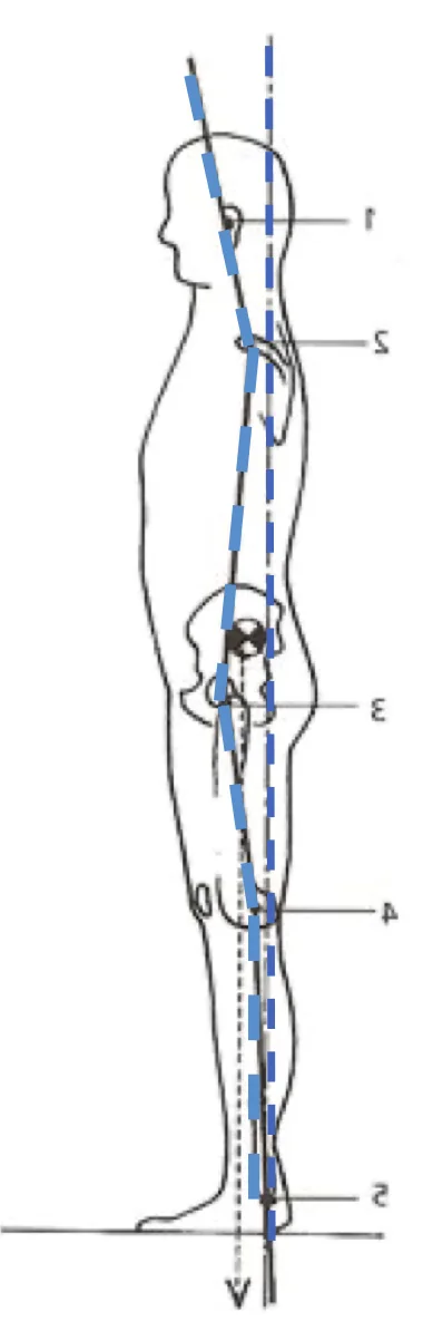
The posture–occlusion interdependence can be objectified in the office using the Meersseman test: a transient occlusal change (e.g., symmetrical shims/cotton rolls in the posterior sectors) is followed by an immediate postural assessment. Reproducible variations support the hypothesis of a functional coupling between the occlusal block and postural control (61). A second test, the Occlusal Kinesthetic Sensitizing Test (OKST) (von Piekartz, 2015), explores the ability to modify neuromuscular signs and symptomatology through an alteration of dental afferents during habitual occlusion. It allows assessment of the sensitivity of the masticatory system to occlusal proprioceptive inputs and their repercussion on craniocervical coordination (88). However, caution is warranted in interpreting these two tests, since the available data do not allow a direct causal relationship to be established between occlusion and posture, the observed correlations possibly reflecting adaptive or compensatory mechanisms (Perinetti 2009). In addition, a third test consists of the objective evaluation of the patient’s global posture, ideally barefoot, with the neck uncovered, in order to observe body symmetry, axial alignment (tragus – acromion – greater trochanter – lateral femoral condyle – lateral malleolus) and any segmental compensations under neutral conditions (60, 70). (Figure 10)
2.4.4 Functional management
Clinical management is organized around three axes:
- Restoring an anatomical environment conducive to function (transverse and sagittal balances, upper airway patency (21)) in order to reduce compensatory tonic stresses.
- Retraining through motor control exercises and muscle strengthening.
- Adapting to the existing craniofacial anatomical constraints and deformities.
2.5 Mastication
2.5.1 Physiology and craniofacial role
Physiological mastication is unilateral alternating, balanced, painless, and mouth-closed. This activation mode symmetrically distributes loads at the TMJs, coordinately recruits the muscles, and participates in craniocervical postural stability. Within this framework, the maxilla receives regular functional shaping, the mandible follows centered kinematics, and neuromuscular tone remains balanced.
Conversely, a reduction in masticatory effort (soft diet) decreases the mechanical stimulation of bone. In accordance with Wolff’s law (90), bone — an anisotropic and viscoelastic material — forms and resorbs according to the stresses it undergoes: fewer stresses result in less transverse shaping of the maxilla and can promote maxillary constriction.
The load-bearing capacities of the masticatory system are considerable: classically, ≈ 10 kgf/cm² of muscle cross-section have been reported, for an average total elevator surface area of ≈ 19.5 cm², i.e., a theoretical force that can approach ≈ 195 kgf during maximal bite. These load levels explain the potential impact of masticatory function on skeletal growth and remodeling. The masticatory system is one of the most powerful muscular systems in the human body and the most finely regulated (62).
2.5.2 Dysfunctions and mechanisms
Habitual non-alternating unilateral chewing constitutes the most common presentation. It may be maintained by a:
- dental pain;
- asymmetry of tone/posture;
- mandibular functional shift.
Biomechanically, the working side concentrates the stresses, while the non-working side is under-recruited; a dysmorpho-functional vicious circle then develops, combining functional shift, rotation of the lower third, and muscular imbalances.
The clinical data confirm this function–structure link. In a comparative study of children with unilateral posterior crossbite, 73 % presented condylar asymmetry greater than 6 % versus 21 % of controls, with a mean asymmetry difference of 10.7 % vs 4.2 % (p < 0.001) (85). This observation supports the hypothesis that a unilateral functional disorder can induce asymmetric bone adaptations.
2.5.3 Clinical and orthodontic consequences
These patterns lead to facial asymmetries and narrow arches, crossbites, functional shifts, as well as asymmetric wear related to non-centered mandibular paths. In the long term, these alterations increase the risk of TMJ dysfunction.
2.5.4 Clinical screening
Screening relies on history-taking (chewing preference, food textures), extra-oral examination (symmetries, opening/closing trajectories, deviation), intra-oral examination (premature contacts, alveolar/maxillary constriction, wear facets) and, if needed, on additional investigations (analysis of the masticatory cycle in real-life conditions). Analysis of mandibular trajectories and of contacts in protrusion/laterotrusion helps to objectify the mandibular functional shift.
2.5.5 Principles of management
The objective is twofold: to restore the anatomical environment conducive to chewing on alternating sides, and then to retrain the function.
- Educational and dietary measures. Informing the patient and parents about the role of mastication in growth, in the formation of the food bolus and in neuromuscular balance; progressive diversification of textures and adaptation of the pace of meals in order to increase masticatory effort from the earliest age.
- Orofacial myofunctional therapy. Exercises of awareness and repeated voluntary training, with natural biofeedback, aimed at (re)establishing an alternating unilateral pattern and at coordinating the action of the elevator muscles with the mandibular trajectory.
- Occlusal or orthopedic corrections. Depending on age and etiology, Planas inclined planes may be used to guide the mandible toward more symmetrical relationships and to stimulate alternating mastication during growth. Transverse expansion (rapid palatal expander, quad-helix) may be indicated in cases of maxillary or alveolar constriction that maintains a mandibular functional shift.
These interventions ideally fit within a multidisciplinary management approach (pediatric dentistry, orthodontics, orofacial myofunctional therapy, dietetics), with monitoring of adherence and clinical progress.
2.6 Tongue posture and swallowing
Recent literature confirms the close interplay between tongue posture, swallowing patterns and craniofacial morphogenesis. In a clinical study of 100 patients, Deregibus et al. reported a higher prevalence of ankyloglossia in Class III subjects as well as an alteration of resting tongue posture in these same groups, suggesting a structure-function coupling that contributes to the antero-posterior expression of malocclusions (19). This relationship is not limited to cross-sectional observations. A systematic review concludes that dysfunctional swallowing significantly and measurably influences malocclusions through tongue position and orofacial neuromuscular balance. To improve occlusal stability, the authors recommend combined management associating orthodontic treatment and orofacial myofunctional therapy, possibly supported by adjunctive oral appliances (42). This dynamic is bidirectional: a narrative review emphasizes that a malocclusion can induce or maintain abnormal swallowing, which, in turn, perpetuates the dentofacial deformity and predisposes to relapse if the function is not retrained in parallel with orthodontic treatment (59). Taken together, these studies justify an integrated strategy that simultaneously targets form (arches, intermaxillary relationships) and function (tongue posture, swallowing praxes).
2.6.1 Definition and physiology
Swallowing is a complex reflex act ensuring the transfer of the bolus (or saliva) from the oral cavity to the stomach. It appears in utero (swallowing of amniotic fluid), then matures during childhood to evolve from an infantile pattern, characterized by marked labial and perioral components, toward an adult pattern. In physiological adult swallowing, the voluntary oral phase is distinguished by elevation of the tongue against the palate — tip resting on the retro-incisal papillae — and by a posterior undulation of the dorsum of the tongue that propels the bolus toward the oropharynx (peristaltic movement). The lips remain at rest, without hypertonicity, while the soft palate rises early and the teeth come briefly into contact. The pharyngeal (reflex) and esophageal (automatic) phases then ensure protection of the airway and progression of the bolus. At rest, the physiological posture is that of a high tongue, whose lateral borders are in contact with the palatal slopes, contributing to transverse stimulation and to the stability of the maxillary architecture. Conversely, the persistence of an infantile pattern beyond the transition age constitutes dysfunctional swallowing, liable to maintain occluso-functional imbalances (59, 42).
2.6.2 Pathophysiology, origins and dysmorpho-functional cascade
The origins are multifactorial:
- Functional:
- mouth breathing keeps the tongue low;
- parafunctions mechanically obstruct tongue elevation;
- habitual (non-alternating) or reduced unilateral chewing impoverishes the lateral and vertical demand on the tongue, weakening its palatal-molding role.
- Anatomical:
- maxillary (transverse) constriction restricts the available palatal space;
- a short lingual frenulum limits elevation;
- a relative macroglossia may hinder palatal adjustment (19, 59, 42);
- orofacial hypotonia makes it more difficult to maintain a high posture.
These factors feed a cascade: a low tongue and dysfunctional swallowing deprive the palate of stimulation, favoring transverse narrowing (often a lyre-shaped arch, a suggestive sign of excessive buccinator muscle contraction), as well as the appearance of crossbites and asymmetries. In addition, tongue thrust contributes to open bites and incisor proclination, and interposition can mimic or maintain a Class II. The literature emphasizes the reciprocal nature of these influences and the fact that a purely morphological treatment predisposes to relapse if the function is not corrected (59, 42).
2.6.3 Screening and management
Screening combines targeted functional history, observation at rest (tongue posture, lip competence, perioral contraction) and in function (anterior thrust, interpositions, tongue mobility/elevation), supplemented, if needed, by analysis of the arches and intermaxillary relationships. Prioritization of care is crucial: restore a favorable ventilatory context and anatomical environment (tongue mobility), then initiate orofacial myofunctional therapy.
From a therapeutic standpoint, a systematic review advocates a combined approach associating orthodontics and myofunctional therapy in order to achieve occlusal stability and to avoid relapses (42). A recent randomized controlled trial in the mixed dentition illustrates the value of this approach in anterior open bite: adding brief orofacial myofunctional therapy to orthodontic treatment attenuates the increase in tongue pressure during swallowing and reduces the persistence of dysfunctional swallowing after appliance removal — a factor favorable to the stability of the result; tongue function is moreover objectively quantified by measuring tongue pressure with an intra-oral dynamometer (50). Passive adjuncts may help to guide treatment as a complement to an active protocol (42).
2.7 Speech
In children, speech sound disorders are accompanied by a higher prevalence and greater severity of malocclusions compared with peers with typical speech development (p < 0.01) (64). A scoping review including 12 studies (more than 2,500 participants) highlights strong and consistent correlations between anterior open bite and difficulties in articulating sibilants (/s/, /z/, /ʃ/), as well as between Class II/III and alterations of /r/, /t/, /d/ (3). All the data converge toward a bidirectional interaction: tongue posture/motricity affect articulation and maxillofacial growth, while dentoskeletal morphology shapes the space available for speech and may perpetuate the articulation disorder (64, 3).
The consonants /t d n l/ require precise anterior elevation of the tongue against the alveolar palate, neutral labial sealing and mandibular stability. Any alteration of these parameters — related to a dysfunction or dentofacial deformity — disrupts articulation, leading to interdentalization or dorsalization phenomena. Recent studies confirm recurrent articulatory distortions in anterior open bites, dorsalization of tongue contacts in high-arched palates, and a low and retropositioned tongue in Class III (5, 33, 45). After orthognathic surgery, alveolar phonemes often remain altered, requiring specific tongue myofunctional therapy (53). Recent reviews emphasize a significant correlation between malocclusion and articulation disorders, particularly for anterior open bite and posterior crossbite (2, 3). These observations reflect a bidirectional interaction between dentoskeletal morphology and tongue motricity: the shape of the arches conditions the space available for speech, while articulatory function influences orofacial growth. Targeted myofunctional therapy of the apico-alveolar contacts is therefore essential to normalize the production of /t d n l/ and to prevent functional relapses.
2.7.1 Definition & physiology
Speech results from the integration of respiration, laryngeal vibration and supralaryngeal articulation. The tongue modulates the resonance cavities and the precision of the points of articulation. A tongue elevated toward the anterior palatal dome stabilizes the airway corridor; conversely, a low or anterior tongue reduces the velolingual space, displaces the contacts and induces labio-mandibular compensations unfavorable to occlusal stability.
2.7.2 Pathophysiology, origin & dysmorpho-functional cascade
The speech disorder may impose an inappropriate lingual pattern (low or anterior rest, repeated contacts) that alters the anterior dental balance, favors open bite and disrupts the incisor relationships. Conversely, a maxillary (transverse) constriction, an anterior open bite or a sagittal discrepancy restrict the space available for speech, impose bypassing trajectories and maintain the articulation disorder (particularly for /s/, /z/, /ʃ/, /r/). This interaction illustrates a dysmorpho-functional cascade justifying joint management of form and function (3).
2.7.3 Screening & management
Screening combines orthodontic and functional examination, observation of resting tongue posture, of contact points during speech and a targeted phonetic analysis (sibilants, apico-alveolars, rhotics). Assessment of co-factors (nasal breathing, frenulum, tonsillar volume) is essential.
Management relies on orofacial myofunctional therapy aimed at lingual stabilization, breath-palate-tongue coordination and the reconstruction of specific articulatory gestures with transfer into spontaneous speech. It will be important to focus myofunctional therapy on a new imprinting of this reconstruction through the repetition of exercises and the patient’s awareness. In parallel, orthodontic treatment corrects the morphological obstacles. Interdisciplinary coordination is recommended to durably break the function-form loop (3, 64).
3Orthodontic clinical situations related to orofacial dysfunctions
3.1 Transverse maxillary deficiency, the common denominator
A narrow palate is the most frequent expression of the dysmorpho-functional cascade. It results from an imbalance between centrifugal forces (tongue) and centripetal forces (cheeks/lips), with two main mechanisms:
- Insufficient centrifugal forces (tongue):
- Related to structures. A restrictive lingual frenulum limits palatal contact and apical elevation of the tongue, decreasing the repeated micropressures of the tongue that allow transverse stimulation of the maxilla and thus favoring alveolar constriction (9, 86).
- Related to postural attitudes. A low tongue posture — frequently associated with unfavorable cervico-cephalic determinants and with tongue–hyoid bone–cervical spine relationships — also reduces the physiological palatal micropressures and contributes to maxillary narrowness (21, 29, 79).
- Related to functions.
- Mouth breathing. The oral air passage keeps the tongue in a low position and consequently impoverishes the repeated palatal stimulations, while depriving the nasal cavity of the morphogenic flow that participates in the widening of the nasal/palatal floor through successive pressure interplay (37, 82, 18, 22, 65).
- Insufficient/weakly tonic mastication. Reduced masticatory demand, often with unilateral patterns, decreases the constructive transverse stresses and contributes to the transverse deficiency (85).
- Parafunctions. Sucking and nibbling habits prevent the rise of the tongue and contribute to maxillary constriction (68, 87, 89).
- Dysfunctional swallowing. The absence of effective palatal contact during swallowing perpetuates the insufficiency of centrifugal forces and the palatal narrowness (59, 42).
- Excess of centripetal forces (cheeks/lips): related to functions. Dysfunctional swallowing (hyperactivity of the buccinator and/or the orbicularis) and/or digit sucking increase the centripetal pressures, shift the neutral zone inward and favor alveolar constriction (59, 17, 87, 89, 68).
Clinically, the transverse deficiency may be:
- manifest (unilateral/bilateral crossbites, lateral edge-to-edge);
- or masked (relative alveolar constriction revealed after repositioning into Class I).
The therapeutic strategy then aims to restore lingual palatal stimulation and to reduce buccal constraints, in keeping with the hierarchy of management.
3.2 Mandibular kinetic adaptations
Faced with a narrow and unstable maxillary arch, the mandible seeks an occlusal path compatible with the neutral zone. Two adaptations are particularly typical:
- Mandibular functional shift: the mandible slides laterally to clear a premature contact. Clinically, one finds a unilateral crossbite, a deviation of the mandibular midline relative to the mid-sagittal plane and, on the side of the shift, Class II relationships. On the opposite side, the relationships are most often Class I (sometimes Class III).
- Class III: the mandible may advance kinetically to bypass an anterior transverse lock (kinetic Class III). The incisor or lateral premature contact then induces an edge-to-edge or an anterior and/or posterior crossbite.
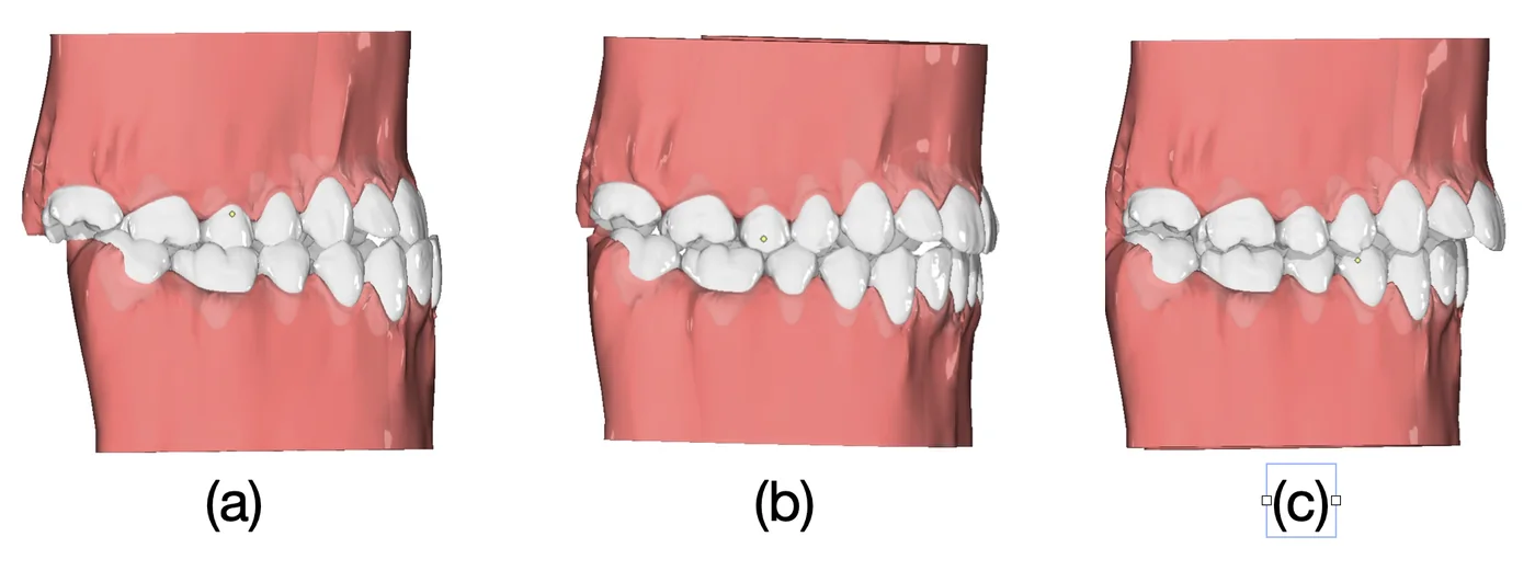
- Class II: the narrow maxilla can act as a transverse lock that blocks mandibular growth and keeps it in a Class II relationship. In this situation, the transverse defect is sometimes visible only in clinical or digital simulation of the antero-posterior correction. Once repositioned into Class I, the patient reveals an edge-to-edge or lateral crossbite occlusion: this is the alveolar constriction relative to Class II, which reflects the underlying transverse discordance masked by the antero-posterior compensation.
3.3 Functional etiologies: the cascade
Several dysfunctions can lead to the same phenotype of transverse maxillary deficiency through converging pathways:
- Oral or mixed breathing: the nasal cavities develop insufficiently, the tongue remains low to let air pass, and the mechanical stimulation of the palate by the tongue disappears. Transverse growth of the maxilla is reduced.
- Restrictive lingual frenulum (ankyloglossia): limitation of tongue elevation, chronic low posture, lack of palatal stimulation and transverse delay.
- Sucking parafunctions (thumb, pacifier): mechanical obstacle to the rise of the tongue and buccinator hyperactivity. The neutral zone contracts, leading to a transverse deficiency.
- Dysfunctional swallowing: the perioral contractions maintain transverse centripetal forces opposed to the expansion forces. The arch takes on a lyre shape, a suggestive (but non-specific) sign of dysfunctional swallowing.
- Mastication: a soft diet reduces the intensity and frequency of the stresses applied to the bone (Wolff’s law), which impoverishes transverse growth; unilateral chewing asymmetrically stimulates the structures and favors deviations.
The whole illustrates the two-way nature of the relationship: function shapes form, but form disrupts function, maintaining the vicious circle.
Beyond the transverse defect, tongue interpositions or thrusts and asymmetries of mastication generate recurrent pictures: anterior and posterior open bites, inter-incisor diastemas from repeated thrusts, and tilting of the occlusal plane related to asymmetrical functional patterns and/or an unbalanced cephalo-cervical posture.
3.4 Management: mechanical and functional
Once the indication is established, maxillary disjunction or maxillary expansion corrects the transverse dimension. However, stability depends on retraining the functions according to a logical sequence:
- Restore nasal breathing to allow lip competence and free up high tongue posture (ENT).
- Remove anatomical and behavioral obstacles: restrictive lingual frenulum, digit or pacifier sucking.
- Retrain resting tongue posture and swallowing: tongue against the palate, tip on the retro-incisal papillae, elimination of anterior tongue thrust and buccinator hyperactivity.
- Restore alternating bilateral mastication of sufficient intensity, in order to maintain the width achieved.
This approach falls within Moss’s functional matrix theory: a correctly positioned tongue, nasal airflow, and effective mastication constitute functional constraints that stabilize the restored skeletal architecture. Without this functional whole, relapse is likely.
3.5 The “wire syndrome”: an apparent relapse with a functional driver
Wire syndrome corresponds to a relapse of functional origin. It takes the form of tooth movements occurring despite a bonded retainer, often after partial adhesive–wire debonding: the tooth loses its anchorage and migrates under the effect of a functional force (tongue thrust/interposition, perioral hyperactivity), while the wire guides the movement, hence the misleading illusion of an “untwisted” wire (13, 78).
Durable correction requires:
- removal of the offending wire;
- neutralization of the dysfunction;
- resumption of treatment if necessary;
- placement of a passive retainer after repositioning, followed by monitoring (4, 13, 43, 78).
3.6 Importance of myofunctional therapy for dysfunctions in a surgical context
In orthognathic surgery, the operation triggers a regional acceleratory phenomenon (RAP) (28, 55) that transiently increases bone remodeling and lowers dento-alveolar resistance: any tooth movement — intended or induced by a dysfunction — can then accelerate. Without orofacial myofunctional therapy, this frequently translates into early and amplified relapses (reopening of the open bite, loss of transverse gain, sagittal drift) (4). Furthermore, regarding stability, the literature reminds us that vertical corrections (including anterior open bite) and certain transverse expansions are among the least stable movements, which reinforces the need for a functional protocol both pre- and, above all, post-operatively (4, 36, 43, 73, 81).
4Discussion
4.1 Current care pathway: referral to myofunctional therapists (gold standard)
The management of dysfunctions relies on referral to trained myofunctional therapists, consistent with international recommendations that integrate the examination and correction of functions as early as possible in the orthodontic pathway (16, 20, 63). The orthodontist remains the conductor of the pathway and can rely on professional directories, but the automation of correct functional patterns requires active and progressive therapy.
4.2 “Passive” myofunctional devices: concepts and role
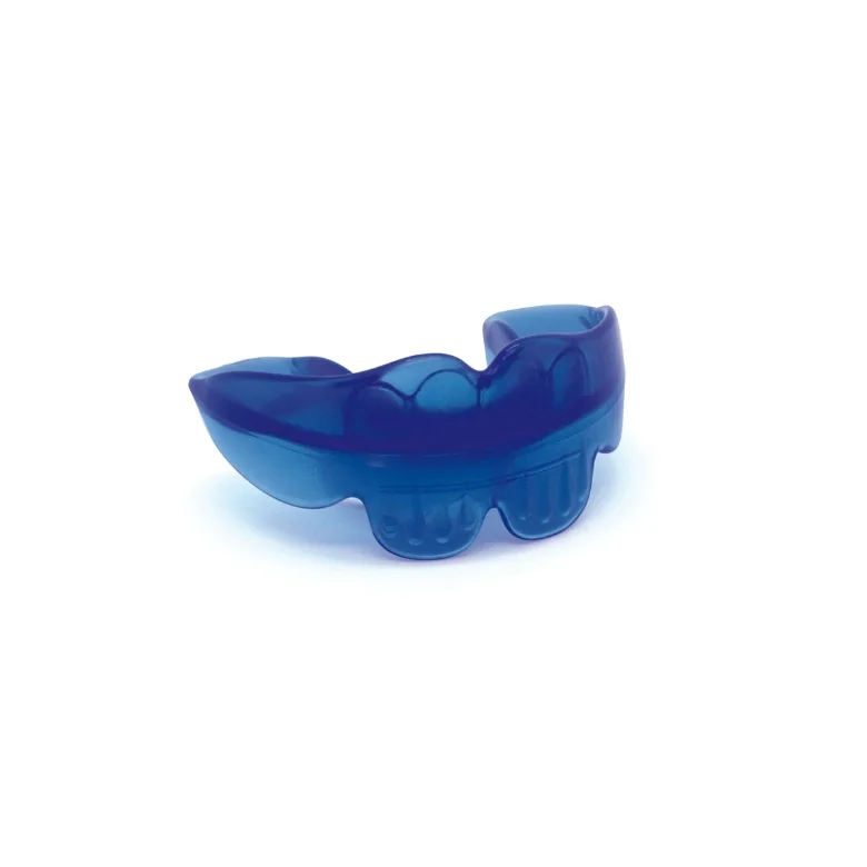
Faced with the difficulties of delivering myofunctional therapy, several devices complement the orthodontist’s therapeutic arsenal. These aim to guide or promote postures/movements and to support active training: functional trainers (Orthoplus®), Myobrace®, FroggyMouth®, Myosimple®, Innovafarm® and the like. Their concept is to offer a geometry and/or labio-linguo-buccal supports that orient the tongue and lips, limit certain dysfunctional contractions, and provide a repeatable proprioceptive reference. These devices can complement active myofunctional therapy but cannot suffice on their own to correct a motor automatism: they do not teach lingual coordination or the contextual automation of functions without explanations or investment from the care team. Their use must therefore be part of an active, planned, and evaluated protocol. Prefabricated myofunctional devices (passive therapy) have only a low level of evidence and do not replace active myofunctional therapy (52).
4.3 Principles of active myofunctional therapy: neuroplasticity and the “10 principles”
Myofunctional therapy relies on experience-dependent neuroplasticity: repeating a movement with intention and attention, relying on biofeedback to make it conscious, in order to achieve the engramming of a new, durable motor pattern. Operationally, Kleim & Jones’s “ten principles” provide a framework for designing progressive, specific, and dosed sequences (frequency × duration × difficulty) (46). Biofeedback — proprioceptive, tactile, visual, or auditory — accelerates acquisition and automation by correcting execution errors in real time and reinforcing awareness of the movement (40). “Passive” appliances can serve as reference points and limit certain compensations, but durable correction is obtained through voluntary, repeated, and structured training consistent with these principles.
4.4 Therapeutic adherence in orofacial myofunctional therapy
Therapeutic adherence — defined as the degree to which a patient’s behavior corresponds to the clinical recommendations provided to them — represents one of the most significant challenges of orofacial myofunctional therapy (OMT). Unlike passive interventions, OMT is by its very nature dependent on active, repeated, and intentional patient participation between sessions. The neurophysiological basis of this requirement is rooted in the principles of experience-dependent neural plasticity: the consolidation of new motor patterns requires sustained repetition, coupled with conscious intention and sensory feedback, to induce durable cortical reorganization (46, 40). In clinical practice, this translates into a minimum of three to six months of daily home exercises to automate functional corrections. In the absence of regular adherence, therapeutic gains remain insufficient to produce lasting neuromuscular change, and the risk of functional relapse after orthodontic treatment is substantially increased (81).
Several barriers to adherence have been identified in the literature on home exercise programs: loss of paper materials, progressive demotivation, insufficient understanding of the therapeutic benefit, logistical constraints related to weekly travel, rigidity of professional and family schedules, and difficulties in accessing care in underserved areas. Data from the general literature confirm these observations: in populations with musculoskeletal pain, only 48.8% of patients fully adhere to the prescribed program, with 27.6% partial adherence and 23.6% non-adherence (71). Comparable results have been reported in other functional therapy contexts, with full adherence rates ranging between 48 and 50% (24). Although no large-scale data specific to OMT are available, these figures are consistent with real-world clinical experience and constitute a recognized obstacle to therapeutic efficacy.
Digital tools have emerged as promising adjuncts to address these structural barriers. A systematic review of ten randomized controlled trials involving 1,117 participants demonstrates that adding a digital intervention to a home exercise program significantly improves adherence in seven of ten trials (49). While the magnitude of long-term effects warrants confirmation, the short-term benefit is well documented and directly applicable to the three-to-six-month time window characteristic of OMT protocols. These tools address the identified barriers in a targeted manner: automated notifications and reminders (forgetting), gamification and reward systems (demotivation), educational video content accessible on replay (misunderstanding), remote access eliminating travel, schedule freedom independent of the clinical agenda, and online submission of exercises replacing paper materials. The integration of a biofeedback mechanism — allowing the patient to receive real-time information on the quality of their motor movement — further reinforces engagement and accelerates motor acquisition through an active feedback loop (40). A multicenter randomized controlled trial protocol (Ibáñez-Rodríguez et al., 2025) specifically evaluating a myofunctional therapy application as an adjunct to improving adherence in the management of OSA validates this approach within the orofacial field (41).
From a clinical governance standpoint, digital monitoring of adherence meets a dual need: for myofunctional therapists, it offers objective documentation of patient engagement that paper-based follow-up could not guarantee; for the orthodontist, it constitutes a structured and documentable record of the functional component of care — a dimension which, as international recommendations remind us, falls within a recognized clinical standard (16, 20, 63).
4.5 Outlook: toward pathways supported by digital technology
Digital tools can remove two major barriers: access to care and documentation. Telemonitoring and interactive education improve adherence in several fields of dentistry and orthodontics (35, 47, 80). Transposed to myofunctional therapy, contextualized reminders, micro-goals, exercise journals, and visual biofeedback at home can increase the “dose–response” necessary for plasticity. In this context, dedicated applications, such as The Tongue Teacher®, can offer exercise programs, educational structuring, planned and hierarchical progression, as well as adherence tracking.

4.6 Current limitations and research perspectives
Despite persistent methodological heterogeneity (variable definitions of mouth breathing, non-standardized morphometric measurements, sometimes short follow-up durations), a body of converging studies supports the integration of functional screening and orofacial myofunctional therapy into the orthodontic pathway (94). The consensus published by Zhou et al. (2024) emphasizes a structured evaluation of dysfunctions, airway analysis, and interdisciplinary coordination. In this context, we propose the SPF pyramid (Structure–Posture–Function) as a clinical methodological framework for organizing, in the orthodontic practice, all the elements to be considered and for guiding the implementation of myofunctional therapy (81, 75). Along these lines, it appears necessary to conduct multicenter controlled trials, with standardized functional criteria and medium- and long-term follow-up, in order to raise the level of evidence and further optimize management protocols.
5Conclusion
Orofacial myofunctional dysfunctions are common and influence orthodontic stability and craniofacial growth (32, 83, 82, 56). Their systematic, documented, and early screening falls within a recognized clinical standard (16, 20, 63). The SPF pyramid provides a clear operative sequence — remove the obstacles (nasal breathing, frenulum, parafunctions), correct the functional environment through orthodontic treatment, then actively retrain tongue posture, swallowing, mastication, and speech — consistent with the functional matrix (Moss) and bone adaptation (Wolff) (65, 90). Combining orthodontics and myofunctional therapy reduces relapses, particularly for anterior open bite (81). Management is interdisciplinary and can rely on digital tools for adherence and documentation; it also enables standardized, reproducible screening that can be deployed on a large scale (35). Multicenter trials with standardized functional criteria remain necessary to raise the level of evidence (94).
6Figure legends
Figure 1. Penfield’s sensorimotor homunculus, illustrating the cortical representation of the different parts of the body, with a marked dominance of the orofacial structures and the hands.
Figure 2. SPF pyramid (structure – posture – function) of orofacial myofunctional determinants, prioritizing therapeutic management.
Figure 3. Diagram of the study by Smithpeter & Covell (2010) comparing relapses in the control group (orthodontics alone) with the experimental group (orthodontics + myofunctional therapy).
Figure 4. Intraoral photograph of a restrictive lingual frenulum.
Figure 5. Representation of the neutral zone, schematizing the main muscular forces present: centripetal (orbicularis oris and buccinator muscles) and centrifugal (tongue muscles).
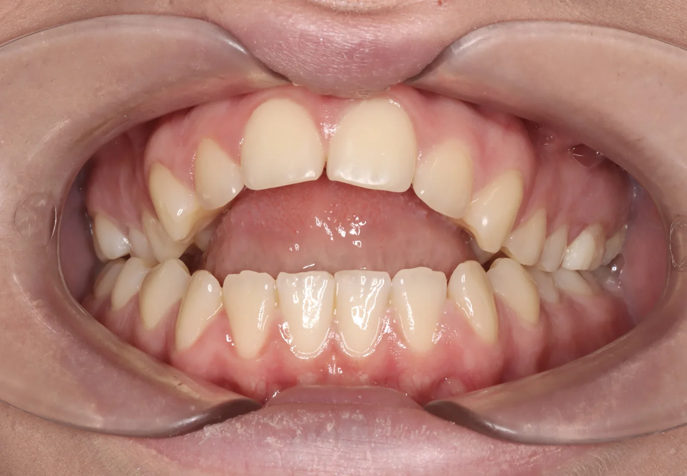
Figure 6. Frontal intraoral photograph objectifying an anterior open bite in a female patient presenting with breathing dysfunction.
Figure 7. Dysmorpho-functional cascade.
Figure 8. Friedman grades for the evaluation of tonsillar volume: Grade 0 — the uvula and the pillars of the tonsillar fossa are visible; Grade 1 — the tonsils are hidden within the fossa; Grade 2 — the tonsils extend beyond the fossa; Grade 3 — the tonsils extend well beyond the fossa without crossing the midline; Grade 4 — the tonsils are in contact at the level of the uvula.
Figure 9. Modified Mallampati score: a tool for evaluating oropharyngeal crowding and the risk of upper airway obstruction. Class 1 — the uvula and the tonsillar fossae are visible; Class 2 — the uvula is partially visible; Class 3 — only the soft palate is visible; Class 4 — only the hard palate is visible.
Figure 10. Landmarks of the tragus–acromion–greater trochanter–lateral femoral condyle–lateral malleolus alignment.
Figure 11. Frontal simulation of a functional lateral shift through mandibular adaptation to a maxillary transverse deficiency: (a) frontal occlusion in lateral edge-to-edge; (b) frontal occlusion in functional lateral shift.
Figure 12. Anteroposterior simulation: (a) Class II with relative alveolar constriction; (b) Class I in transverse edge-to-edge; (c) functional Class III; positioning in Class I if the patient is in Class II, or in centric relation in the case of a Class III occlusion, reveals the transverse deficiency and the edge-to-edge occlusion, as well as the skeletal or kinetic adaptation.
Figure 13. Functional trainer.
Figure 14. Screenshot of the tongueteacher.com website displaying the dashboard and The Tongue Teacher application, regarding patient adherence tracking detected with the help of artificial intelligence, according to the different programs.
7References
- Abtahi S, Witmans M, Alsufyani NA, Major MP, Major PW. Pediatric sleep-disordered breathing in the orthodontic population: prevalence of positive risk and associations. Am J Orthod Dentofacial Orthop. 2020;157(4):466–473.e1.
- Amr-Rey O, et al. Association between malocclusion and articulation disorders: a multicriteria analysis. Eur J Orthod. 2022;44(6):621–628.
- Aprile M, Verdecchia A, Dettori C, Spinas E. Malocclusion and its relationship with sound speech disorders in deciduous and mixed dentition: a scoping review. Dent J (Basel). 2025;13(1):27.
- Arqub SA, et al. The dark side of fixed retainers: Case series of unintended tooth movement. Am J Orthod Dentofacial Orthop. 2023;164(6).
- Assaf D do C, et al. Association between malocclusion, tongue position and speech distortion in children. Braz Oral Res. 2021;35:e112.
- Baxter R, Zaghi S, Kaplan M, Montes M, Guilleminault C. Functional definition of restrictive tongue-tie (ankyloglossia) with implications for frenuloplasty and myofunctional therapy. Int J Pediatr Otorhinolaryngol. 2020;128:109697.
- Bedoucha V, Boutin F, Frapier L. Impact of genioplasty during puberty on the upper airways. Int Orthod. 2015;13(4):421–435.
- Behrents RG, Shelgikar AV, Conley RS, Flores-Mir C, Hans M, Levine M, et al. Obstructive sleep apnea and orthodontics: An American Association of Orthodontists White Paper. Am J Orthod Dentofacial Orthop. 2019;156(1):13–28.e1.
- Calvo-Henríquez C, Martins Neves S, Branco AM, Lechien JR, Betances Reinoso F, Mota Rojas X, et al. Relationship between short lingual frenulum and malocclusion: a multicentre study. Acta Otorrinolaringol Esp (Engl Ed). 2022;73(3):177–183.
- Camacho M, Certal V, Abdullatif J, Zaghi S, Ruoff C, Capasso R, et al. Orofacial myofunctional therapy for obstructive sleep apnea: a systematic review and meta-analysis. J Clin Sleep Med. 2023;19(9):1611–1623.
- Cammaroto G, Galletti F, Calvo-Henriquez C, Vicini C, Montevecchi F. Effectiveness of myofunctional therapy to treat adult obstructive sleep apnea: an updated systematic review. J Clin Med. 2024;13(4):1179.
- Chamberland S, Proffit WR, Chamberland P-E. Functional genioplasty in growing patients. Angle Orthod. 2015;85(3):360–373.
- Charavet C, Picon A, Manière-Ezvan A, d’Incamps O. “Wire Syndrome” Following Bonded Orthodontic Retainers: A Systematic Review. J Clin Med. 2022;11(5):1363.
- Chervin RD, Archbold KH, Panahi P, Pituch KJ. Pediatric sleep questionnaire (PSQ): validity and reliability. Sleep Med. 2000;1(1):21–32.
- Cohen-Lévy J, Vion A, Lobre C, Pétel V, Fleury B. The influence of obstructive sleep apnea syndrome on orthodontic treatment decisions regarding children and adolescents. Int Orthod. 2023;21(2):100746.
- Consejo General de Dentistas de España / Fundación Dental Española (FDE). La salud bucodental en España: ¿dónde estamos y hacia dónde vamos? Madrid.
- Cozza P, Baccetti T, Franchi L, Mucedero M. Transverse features of subjects with sucking habits and facial hyperdivergency in the mixed dentition. Am J Orthod Dentofacial Orthop. 2007;132(2):226–229.
- Dallam G, Kies B. The effect of nasal breathing versus oral and oronasal breathing during exercise: a review. J Sports Res. 2020;7(1):1–10.
- Deregibus A, Castroflorio T, Debernardi C, Trovato FM, Bonetti GA. Analysis of tongue function from the orthodontist’s point of view: Not only a matter of deglutition. Appl Sci. 2021;11(1):160.
- Deutsche Gesellschaft für Kieferorthopädie (DGKFO), DGZMK. S2k-Leitlinie: Ideale Zeitpunkte und Maßnahmen der kieferorthopädischen Diagnostik. AWMF 083-050. Juni 2025.
- Dipalma G, Inchingolo AD, Pezzolla C, Sardano R, Trilli I, Di Venere D, et al. Head and cervical posture in sagittal skeletal malocclusions: insights from a systematic review. J Clin Med. 2025;14(8):2626.
- Eser P, Calamai P, Simader E, et al. Improved exercise ventilatory efficiency with nasal compared to oral breathing in cardiac patients. Front Physiol. 2024;15:1380562.
- Fernandes Fagundes NC, Loliencar P, MacLean JE, Flores-Mir C, Heo G. Characterization of craniofacial-based clinical phenotypes in children with suspected obstructive sleep apnea. J Clin Sleep Med. 2023;19(11):1857–1865.
- Francisco IM, Tozzo MC, Martins J, de Oliveira AS. Adherence of individuals with shoulder pain to home exercise booklets. Musculoskelet Sci Pract. 2024;72:102956.
- Frapier L, Jaussent A, Yachouh J, Goudot P, Dauvilliers Y, Picot M-C. Impact of genioplasty on mandibular growth during puberty. Int Orthod. 2010;8(4):342–359.
- Frapier L, Picot M-C, Dauvilliers Y, et al. Ventilatory disorders and facial growth: benefits of early genioplasty. Int Orthod. 2011;9(1):20–41.
- Friedman M, Ibrahim H, Bass L. Clinical staging for sleep-disordered breathing. Otolaryngol Head Neck Surg. 2002;127(1):13–21.
- Frost HM. The regional acceleratory phenomenon: a review. Henry Ford Hosp Med J. 1983;31(1):3–9.
- Giannakopoulos NN, Hellmann D, Schmitter M, Krüger B, Hauser T, Hugger A. Anatomical and functional relationships between the hyoid bone, tongue position, and cervical posture. J Oral Rehabil. 2013;40(5):347–353.
- Gonzalez I, Garcia-Guevara H, Viamonte MD, Jensen D, Hernandez A. Changes in the Airway after Mentoplasty (Genioplasty): Systematic Review. Int J Morphol. 2022;40(4):1025–1034.
- Gozal D. Sleep-disordered breathing and school performance/neurocognition in children. Sleep Med Rev. 2004;8(6):447–459.
- Grabowski R, Kundt G, Stahl F. Interrelation between occlusal findings and orofacial myofunctional status in primary and mixed dentition. Part III. J Orofac Orthop. 2007;68(6):462–476.
- Guay AH. Tongue posture at rest and during phonation in Class III malocclusion. Am J Orthod. 1978;74(3):295–300.
- Guilleminault C, Huang YS, Monteyrol PJ, Sato R, Quo S, Lin CH. Critical role of myofascial reeducation in pediatric sleep-disordered breathing. Sleep Med. 2013;14(6):518–525.
- Gurgel-Juarez N, Torres-Pereira C, Haddad AE, et al. Accuracy and effectiveness of teledentistry: a systematic review of systematic reviews. Evid Based Dent. 2022;23(3):100–102.
- Haas Junior OL, Guijarro-Martínez R, et al. Hierarchy of surgical stability in orthognathic surgery: overview of systematic reviews. Int J Oral Maxillofac Surg. 2019.
- Harvold EP, Tomer BS, Vargervik K, Chierici G. Primate experiments on oral respiration. Am J Orthod. 1981;79(4):359–372.
- Hill RR, Lee CS, Pados BF. The prevalence of ankyloglossia in children aged <1 year: a systematic review and meta-analysis. Pediatr Res. 2021;90(2):259–266.
- Hu J, Zhou C, Li J, Zhao H, Wang Z, Zhang J. Efficacy of myofunctional therapy for obstructive sleep apnea: a network meta-analysis. Sleep Breath. 2025;29(1):145–156.
- Huang HC, Wolf SL, He J. Recent developments in biofeedback for neuromotor rehabilitation. J Neuroeng Rehabil. 2006;3:11.
- Ibáñez-Rodríguez JA, O’Connor-Reina C, Lucas R, Rodriguez-Alcala L, Correa E, Ignacio JM, et al. App-based myofunctional therapy to improve adherence and outcomes of mandibular advancement devices in moderate OSA: Multicenter randomized trial protocol. JMIR Res Protoc. 2025;14:e80521.
- Inchingolo AD, Carpentiere V, Inchingolo AM, de Ruvo E, Campanelli M, Ferrante L, et al. Orthodontic treatment in patients with atypical swallowing and malocclusion: a systematic review. J Clin Pediatr Dent. 2024;48(5):14–26.
- Jedliński M, Tandecka K, Grocholewicz K, Janiszewska-Olszowska J. Inadvertent Tooth Movement from a Bonded Mandibular Lingual Retainer—Case Report with 3D Follow-Up and Microscopy. Appl Sci. 2024;14(9):3889.
- Johns MW. A new method for measuring daytime sleepiness: The Epworth Sleepiness Scale. Sleep. 1991;14(6):540–545.
- Keyser MMB, et al. Impacts of Skeletal Anterior Open Bite Malocclusion on Speech. J Pers Med. 2022;12(9):1457.
- Kleim JA, Jones TA. Principles of experience-dependent neural plasticity: Implications for rehabilitation after brain damage. J Speech Lang Hear Res. 2008;51(Suppl 2):S225–S239.
- Korean Association of Orthodontists. Remote digital monitoring during orthodontic retention: a prospective feasibility study. Korean J Orthod. 2022;52(2):123–131.
- Kotlow LA. Ankyloglossia (tongue-tie): a diagnostic and treatment quandary. Quintessence Int. 1999;30(4):259–262.
- Lang S, McLelland C, MacDonald D, Hamilton DF. Do digital interventions increase adherence to home exercise rehabilitation? A systematic review of RCTs. Arch Physiother. 2022;12:24.
- Leal TY, Garib D, Eto H, Rosa R, Miranda F, Soares A, Fukushiro AP, Berretín-Félix G. Brief orofacial myofunctional therapy and tongue pressure as prediction factors for open bite correction: a randomized clinical trial. Clin Oral Investig. 2025;29(9):407.
- Lee EJ, Cho JH. Meta-analysis of obstruction site observed with drug-induced sleep endoscopy in patients with obstructive sleep apnea. Laryngoscope. 2019;129(5):1235–1243.
- Levrini L, Salone GS, Ramirez-Yanez GO. Pre-fabricated myofunctional appliance for the treatment of mild to moderate pediatric obstructive sleep apnea: a preliminary report. J Clin Pediatr Dent. 2018;42(3):236–239.
- Lichnowska A, et al. Logopedic evaluation after orthognathic surgery: recovery of alveolar consonants and role of myofunctional re-education. Appl Sci. 2021;11(14):6563.
- Lin L, Zhao T, Qin D, Hua F, He H. The impact of mouth breathing on dentofacial development: a concise review. Front Public Health. 2022;10:929165.
- Liou EJW. Postoperative rapid orthodontic tooth movement (PRTM). J Oral Maxillofac Surg. 2011;69.
- Malara P, Wozniak K, Szyszka-Sommers K, et al. Outcomes and Stability of Anterior Open Bite Treatment: A Systematic Review and Meta-Analysis. J Clin Med. 2021;10(23):5682.
- Mallampati SR, Gatt SP, Gugino LD, Desai SP, Waraksa B, Freiberger D, et al. A clinical sign to predict difficult tracheal intubation: A prospective study. Can Anaesth Soc J. 1985;32(4):429–434.
- Marcus CL, Brooks LJ, Draper KA, Gozal D, Halbower AC, Jones J, et al. Diagnosis and management of childhood obstructive sleep apnea syndrome. Pediatrics. 2012;130(3):576–584.
- Maspero C, Giannini L, Galbiati G, Farronato G. Atypical swallowing: A review. Minerva Stomatol. 2014;63(5):217–227.
- Maurer-Grubinger C, Adjami F, Ohlendorf D, et al. Symmetrical dental occlusion blocking – changes of body sway and weight distribution in healthy subjects across four age decades. BMC Musculoskelet Disord. 2021;22.
- Meersseman A. Occlusal proprioception and postural control. Cranio. 1999;17(3):189–195.
- Merema MR, van der Bilt A, Weijenberg RAF. Measurement of human bite force: A narrative review. J Oral Rehabil. 2022;49(12):1171–1180.
- Ministero della Salute. Raccomandazioni cliniche in odontostomatologia. Roma; settembre 2017.
- Mogren K, Westerlund S, Bratel J, Dahllöf G. Children and adolescents with speech sound disorders are more likely to have malocclusion. Clin Exp Dent Res. 2022.
- Moss ML, Rankow RM. The role of the functional matrix in mandibular growth. Angle Orthod. 1968;38(2):95–103.
- Nammour S, et al. Laser-assisted lingual frenectomy: functional benefits and orthodontic implications. Lasers Med Sci. 2019;34(6):1121–1128.
- Palomo JM, Cohen-Lévy J, Flores-Mir C, Khosravi R, Levine M, Pickard M, et al. Sleep-disordered breathing and orthodontics: An AAO white paper update. Am J Orthod Dentofacial Orthop. 2026;169(4):419–427.
- Paolantonio EG, Ludovici N, Saccomanno S, La Torre G, Grippaudo C. Association between oral habits, mouth breathing and malocclusion in Italian preschoolers. Eur J Paediatr Dent. 2019;20(3):204–208.
- Penfield W, Rasmussen T. The Cerebral Cortex of Man: A Clinical Study of Localization of Function. New York: Macmillan; 1950.
- Perinetti G. Dental occlusion and body posture: no detectable correlation. Gait Posture. 2009;29(3):494–498.
- Petrosyan H, Leonardi C, Thakral A, Roth J, Russoniello N, Goldin Y, Parikh S. Barriers and factors associated with adherence to a home exercise program of adults with musculoskeletal pain. J Back Musculoskelet Rehabil. 2024;37(2):473–485.
- Poncin W, Willemsens A, Gely L, Contal O. Assessment and rehabilitation of tongue motor skills with myofunctional therapy in obstructive sleep apnea: a systematic review and meta-analysis. J Clin Sleep Med. 2024;20(9):1535–1549.
- Proffit WR, Turvey TA, Phillips C. The hierarchy of stability and predictability in orthognathic surgery with rigid fixation: an update and extension. Head Face Med. 2007;3:21.
- Puhan MA, Suarez A, Lo Cascio C, Zahn A, Heitz M, Braendli O. Didgeridoo playing as alternative treatment for obstructive sleep apnoea syndrome: RCT. BMJ. 2006;332(7536):266–270.
- Ridder G. Zahnstellung und Haltung. München: Elsevier Urban & Fischer; 2016.
- Rocabado M. The hyoid bone position in relation to craniocervical posture. Cranio. 1983;1(3):190–195.
- Rogers AP. Evolution, development, and application of myofunctional therapy in orthodontics. Am J Orthod Oral Surg. 1939;25(1):1–19.
- Roussarie F, Maizeray R, Courtois B, et al. Effet indésirable des fils de contention collés : le « syndrome du fil ». Orthod Fr (JDAO/ODF). 2018;89(4).
- Różańska-Perlińska D, Potocka-Mitan M, Rydzik Ł, Lipińska P, Perliński J, Javdaneh N, Jaszczur-Nowicki J. The correlation between malocclusion and body posture and cervical vertebral, podal system, and gait parameters in children: A systematic review. J Clin Med. 2024;13(12):3463.
- Sangalli L, Savoldi A, Dalessandri D, et al. Effectiveness of remote digital monitoring for improving oral hygiene in orthodontic patients: a prospective study. BMC Oral Health. 2021;21:435.
- Smithpeter J, Covell DA Jr. Relapse of anterior open bites treated with orthodontic appliances with and without orofacial myofunctional therapy. Am J Orthod Dentofacial Orthop. 2010;137(5):605–614.
- Souki BQ, Pimenta GB, Franco LP, Becker HM, Pinto JA. Prevalence of malocclusion among mouth breathing children: Do expectations meet reality? Am J Orthod Dentofacial Orthop. 2009;136(5):e1–e9.
- Stahl F, Grabowski R, Gaebel M, Kundt G. Relationship between occlusal findings and orofacial myofunctional status in primary and mixed dentition. Part II: prevalence of orofacial dysfunctions. J Orofac Orthop. 2007;68(2):74–90.
- Tecco S, Baldini A, Mummolo S, Marchetti E, Giuca MR, Marzo G, et al. Frenulectomy of the tongue and associated myofunctional therapy improve tongue posture and swallowing pattern in subjects with short lingual frenulum. Clin Exp Dent Res. 2015;1(1):6–12.
- Tortarolo A, Rotolo R, Nucci L, Caruso S, Matarese G, Nucera R, et al. Condylar asymmetry in children with unilateral posterior crossbite malocclusion: A comparative cross-sectional study. Children (Basel). 2022;9(11):1772.
- Vaz AC, Bai PM. Lingual frenulum and malocclusion: An overlooked tissue or a minor issue. Indian J Dent Res. 2015;26(5):488–492.
- Viggiano D, Fasano G, Monaco G, Strohmenger L. Breast feeding, bottle feeding, and non-nutritive sucking; effects on occlusion in deciduous dentition. Arch Dis Child. 2004;89:1121–1123.
- von Piekartz P. The occlusal kinesthetic sensitizing test (OKST): a clinical tool to assess sensorimotor integration between dental occlusion and craniocervical posture. J Man Manip Ther. 2015;23(4):199–208.
- Warren JJ, Slayton RL, Bishara SE, et al. Effects of nonnutritive sucking habits on occlusal characteristics in the mixed dentition. Pediatr Dent. 2005;27:445–450.
- Wolff J. Das Gesetz der Transformation der Knochen. Berlin: A. Hirschwald; 1892.
- Xie X, Li X, Zhang L, Liu Y, Chen Y, Li C, et al. Efficiency of orofacial myofunctional therapy in treating obstructive sleep apnea: a systematic review and meta-analysis. Sleep Med. 2022;91:21–29.
- Zaghi S, Valcu-Pinkerton S, Jabara M, Norouz-Knutsen L, Govardhan C, Moeller J, et al. Lingual frenuloplasty with myofunctional therapy: exploring safety and efficacy in 348 cases. Laryngoscope Investig Otolaryngol. 2019;4(5):489–496.
- Zaghi S, Cammaroto G, Valcu-Pinkerton S, Zolty P, Sorrenti G, Hockel M, et al. Functional lingual frenuloplasty with myofunctional therapy: results from an international multi-center cohort study. Cranio. 2023;41(4):336–345.
- Zhou C, Duan P, He H, Song J, Hu M, Liu Y, et al. Expert consensus on pediatric orthodontic therapies of malocclusions in children. Int J Oral Sci. 2024;16(1):32.